Neuroimaging is the set of techniques to image the structure and function of the brain. In this project we collaborate with psychologists and clinical doctors working in several clinical and technical problems. Our first works deal with Attention-Deficit/Hyperactivity Disorder (ADHD) diagnosis using anatomical/structural and functional Magnetic Resonance Images (MRI).
Structural MRI is a medical imaging technique used in neuroradiology to visualize the brain internally in detail.
ADHD is a developmental disorder characterized by: Inattentiveness, Motor hyperactivity, and Impulsiveness. It is the most prevalent psychiatric disorder in childhood and the diagnosis is difficult. There are contradictory studies pointing to overdiagnosis, underdiagnosis, and undertreatment of ADHD.
Neuroanatomical Abnormalities in ADHD:
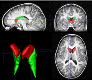
Studies of volumetric brain MRI showed neuroanatomical abnormalities in pediatric ADHD. One of the most replicated findings is the diminished volume of the right caudate nucleus. New diagnostic test based on a ratio between head and body of the caudate have been presented showing statistically different in ADHD and control children. In general, previous studies on ADHD are based on manual procedure, which is extremely time consuming, and prone to inter-rater discrepancies, limiting the power of the analysis.
Automatic ADHD Diagnostic Test:
Thus, there is interest in: 1) Developing automatic methods for segmentation and diagnostic test to accelerate the analysis and make the procedure feasible for large amounts of data. 2) Finding new biomarkers to ADHD diagnosis using MRI which could be useful for clinical practice.
In [Igual2011, Igual2012a, Igual2012b] we introduce a fully automatic diagnostic imaging test for ADHD diagnosis assistance based on previously found evidences of caudate nucleus volumetric abnormalities. The proposed method consists of different steps: a new automatic method for external and internal segmentation of caudate based on machine learning methodologies; the definition of a set of new volume relation features used for caudate representation and classification. We separately validate the contributions using real data from a pediatric population. We present precise internal caudate segmentation results and good discrimination power of the diagnostic test, showing significant performance improvements in comparison to other state-of-the-art methods.
This work was developed in collaboration with the research group in cognitive neurosciences from Universitat Autònoma de Barcelona.
Functional MRI (fMRI) measures brain activity by detecting changes in blood flow. It is used to analyse neural activity during different time-points in a task or resting.
Resting-State fMRI (RS-fMRI) is a brain imaging technique useful for exploring functional connectivity. A major point of interest in RS-fMRI analysis is to isolate connectivity patterns characterising disorders such as for instance ADHD. Such characterisation is usually performed in two steps: first, all connectivity patterns in the data are extracted by means of Independent Component Analysis (ICA); second, standard statistical tests are performed over the extracted patterns to find differences between control and clinical groups. In [Tabas2014], we introduce a novel, single-step, approach for this problem termed Spatial Discriminant ICA. The algorithm can efficiently isolate networks of functional connectivity characterising a clinical group by combining ICA and a new variant of the Fisher’s Linear Discriminant also introduced in this work. As the characterization is carried out in a single step, it potentially provides for a richer characterisation of inter-class differences. The algorithm is tested using synthetic and real fMRI data, showing promising results in both experiments.
RS-fMRI can reveal information of functional connectivity between distant regions during rest. In [Nuñez2015], we present a novel method, called Functional-Anatomical Discriminative Regions (FADR), for selecting a discriminative subset of functional-anatomical regions of the brain in order to characterize functional connectivity abnormalities in mental disorders. FADR integrates Independent Component Analysis with a sparse feature selection strategy, namely Elastic Net, in a supervised framework to extract a new sparse representation. In particular, ICA is used for obtaining group Resting State Networks and functional information is extracted from the subject-specific spatial maps. Anatomical information is incorporated to localize the discriminative regions. Thus, functional-anatomical information is combined in the new descriptor, which characterizes areas of different networks and carries discriminative power. Experimental results on the public database ADHD-200 validate the method being able to automatically extract discriminative areas and extending results from previous studies. The classification ability is evaluated showing that our method performs better than the average of the teams in the ADHD-200 Global Competition while giving relevant information about the disease by selecting the most discriminative regions at the same time.
References:
[Igual2011] L. Igual, J. C. Soliva, A. Hernández-Vela, S. Escalera, X. Jiménez, O. Vilarroyaand P. Radeva. A Fully-Automatic Caudate Nucleus Segmentation of Brain MRI: Application in Volumetric Analysis of Pediatric Attention-Deficit Hyperactivity Disorder, BioMedical EngineeringOnLine 2011, 10:105.
[Igual2012a] L Igual, J. C. Soliva, R. Gimeno, S. Escalera, O. Vilarroya, and P. Radeva. Automatic Internal Segmentation of Caudate Nucleus for Diagnosis of Attention-Deffcit/Hyperactivity Disorder. International Conference on Image Analysis and Recognition (ICIAR) 2012. 25-27|06|2012, Aveiro (Portugal)
[Igual2012b] L. Igual, Joan C. Soliva, S. Escalera, R. Gimeno, O. Vilarroya, P. Radeva. Automatic Brain Caudate Nuclei Segmentation and Classification in Diagnostic of Attention-Deficit/Hyperactivity Disorder. Computerized Medical Imaging and Graphics, 36(8), pp.591-600, 2012. doi:10.1016/j.compmedimag.2012.08.002.
[Nuñez2015] M. Nuñez-Garcia, S. Simpraga, M. A. Jurado, M. Garolera, R. Pueyo, and L. Igual. FADR: Functional-Anatomical Discriminative Regions for rest fMRI Characterization. 6th International Workshop on Machine Learning in Medical Imaging, MLMI in conjunction with MICCAI, 2015.
[Tabas2014] A. Tabas, E. Balaguer-Ballester, L. Igual. Spatial Discriminant ICA for RS-fMRI Characterisation. 4th International Workshop on Pattern Recognition in Neuroimaging, PRNI 2014.
Last update: 01/02/2016.