
Neuroimaging Processing and Analysis
In this project we focus on the processing and analysis of neuroimaging. Neuroimaging is the set of techniques to image the structure and function of the brain. We collaborate with psychologists and clinical doctors working in several clinical and technical problems.
Our first works deal with Attention-Deficit/Hyperactivity Disorder (ADHD) diagnosis using anatomical/structural and functional Magnetic Resonance Images (MRI).
Structural MRI is a medical imaging technique used in neuroradiology to visualize the brain internally in detail.
ADHD is a developmental disorder characterized by: Inattentiveness, Motor hyperactivity, and Impulsiveness. It is the most prevalent psychiatric disorder in childhood and the diagnosis is difficult. There are contradictory studies pointing to overdiagnosis, underdiagnosis, and undertreatment of ADHD.
Neuroanatomical Abnormalities in ADHD
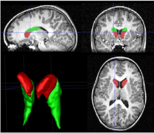
Studies of volumetric brain MRI showed neuroanatomical abnormalities in pediatric ADHD. One of the most replicated findings is the diminished volume of the right caudate nucleus. New diagnostic test based on a ratio between head and body of the caudate have been presented showing statistically different in ADHD and control children.
In general, previous studies on ADHD are based on manual procedure, which is extremely time consuming, and prone to inter-rater discrepancies, limiting the power of the analysis.
Automatic ADHD Diagnostic Test
Thus, there is interest in: 1) Developing automatic methods for segmentation and diagnostic test to accelerate the analysis and make the procedure feasible for large amounts of data. 2) Finding new biomarkers to ADHD diagnosis using MRI which could be useful for clinical practice.
In [Igual2011, Igual2012a, Igual2012b] we introduce a fully automatic diagnostic imaging test for ADHD diagnosis assistance based on previously found evidences of caudate nucleus volumetric abnormalities. The proposed method consists of different steps: a new automatic method for external and internal segmentation of caudate based on machine learning methodologies; the definition of a set of new volume relation features used for caudate representation and classification. We separately validate the contributions using real data from a pediatric population. We present precise internal caudate segmentation results and good discrimination power of the diagnostic test, showing significant performance improvements in comparison to other state-of-the-art methods.
This work was developed in collaboration with the research group in cognitive neurosciences from Universitat Autònoma de Barcelona.
Functional MRI (fMRI) measures brain activity by detecting changes in blood flow. It is used to analyse neural activity during different time-points in a task or resting.
Resting-State fMRI (RS-fMRI) is a brain imaging technique useful for exploring functional connectivity. A major point of interest in RS-fMRI analysis is to isolate connectivity patterns characterising disorders such as for instance ADHD. Such characterisation is usually performed in two steps: first, all connectivity patterns in the data are extracted by means of Independent Component Analysis (ICA); second, standard statistical tests are performed over the extracted patterns to find differences between control and clinical groups. In [Tabas2014], we introduce a novel, single-step, approach for this problem termed Spatial Discriminant ICA. The algorithm can efficiently isolate networks of functional connectivity characterising a clinical group by combining ICA and a new variant of the Fisher’s Linear Discriminant also introduced in this work. As the characterization is carried out in a single step, it potentially provides for a richer characterisation of inter-class differences. The algorithm is tested using synthetic and real fMRI data, showing promising results in both experiments.
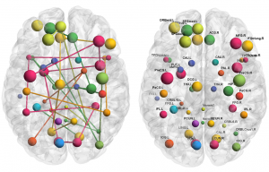
RS-fMRI can reveal information of functional connectivity between distant regions during rest. In [Nuñez2015], we present a novel method, called Functional-Anatomical Discriminative Regions (FADR), for selecting a discriminative subset of functional-anatomical regions of the brain in order to characterize functional connectivity abnormalities in mental disorders. FADR integrates Independent Component Analysis with a sparse feature selection strategy, namely Elastic Net, in a supervised framework to extract a new sparse representation. In particular, ICA is used for obtaining group Resting State Networks and functional information is extracted from the subject-specific spatial maps. Anatomical information is incorporated to localize the discriminative regions. Thus, functional-anatomical information is combined in the new descriptor, which characterizes areas of different networks and carries discriminative power. Experimental results on the public database ADHD-200 validate the method being able to automatically extract discriminative areas and extending results from previous studies. The classification ability is evaluated showing that our method performs better than the average of the teams in the ADHD-200 Global Competition while giving relevant information about the disease by selecting the most discriminative regions at the same time.
References:
[Igual2011] L. Igual, J. C. Soliva, A. Hernández-Vela, S. Escalera, X. Jiménez, O. Vilarroyaand P. Radeva. A Fully-Automatic Caudate Nucleus Segmentation of Brain MRI: Application in Volumetric Analysis of Pediatric Attention-Deficit Hyperactivity Disorder, BioMedical EngineeringOnLine 2011, 10:105.
[Igual2012a] L Igual, J. C. Soliva, R. Gimeno, S. Escalera, O. Vilarroya, and P. Radeva. Automatic Internal Segmentation of Caudate Nucleus for Diagnosis of Attention-Deffcit/Hyperactivity Disorder. International Conference on Image Analysis and Recognition (ICIAR) 2012. 25-27|06|2012, Aveiro (Portugal)
[Igual2012b] L. Igual, Joan C. Soliva, S. Escalera, R. Gimeno, O. Vilarroya, P. Radeva. Automatic Brain Caudate Nuclei Segmentation and Classification in Diagnostic of Attention-Deficit/Hyperactivity Disorder. Computerized Medical Imaging and Graphics, 36(8), pp.591-600, 2012. doi:10.1016/j.compmedimag.2012.08.002.
[Nuñez2015] M. Nuñez-Garcia, S. Simpraga, M. A. Jurado, M. Garolera, R. Pueyo, and L. Igual. FADR: Functional-Anatomical Discriminative Regions for rest fMRI Characterization. 6th International Workshop on Machine Learning in Medical Imaging, MLMI in conjunction with MICCAI, 2015.
[Tabas2014] A. Tabas, E. Balaguer-Ballester, L. Igual. Spatial Discriminant ICA for RS-fMRI Characterisation. 4th International Workshop on Pattern Recognition in Neuroimaging, PRNI 2014.
Automatic Detection and Characterization of Atherosclerotic Plaque in Carotid Ultrasound
Cardiovascular diseases (CVD) are the leading cause of death in Western countries. The common basis of this group of diseases is atherosclerosis, a chronic degenerative process characterized morphologically by an asymmetric focal thickening of the innermost layer of the artery. The carotid arteries may develop atherosclerosis so that blood flow may be partially or totally blocked by the atherosclerotic plaque. The atherosclerotic plaque can be indirectly detected using carotid artery images. In this project, we have proposed a fully-automatic method to detect the presence of plaque in the far wall common carotid artery image [Zhang2015] and we are working in the extension of this method to other segments of the carotid.
Characterization of carotid plaque composition, more specifically the amount of lipid core, fibrous tissue, and calcified tissue, is an important task for the identification of plaques that are prone to rupture, and thus for early risk estimation of cardiovascular and cerebrovascular events. Due to its low costs and wide availability, carotid ultrasound has the potential to become the modality of choice for plaque characterization in clinical practice. However, its significant image noise, coupled with the small size of the plaques and their complex appearance, makes it difficult for automated techniques to discriminate between the different plaque constituents.
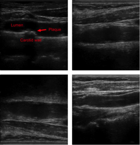
Figure: Four examples of carotid ultrasound images, incorporating noise, artifacts, shadowing, and reverberation. All of these examples contain plaques, but their detection and characterization is challenging even for an expert clinician, resulting in tedious and inconsistent assessments.
In this project, we have addressed this challenging problem by exploiting the unique capabilities of the emerging deep learning framework. More specifically, and unlike existing works, which require a priori definition of specific imaging features or thresholding values, we propose to build a convolutional neural network (CNN) that will automatically extract from the images the information that is optimal for the identification of the different plaque constituents [Lekadir2016]. Based on our previous results, our goal is to progressively build a larger database of training cases together with our clinical partner to further enrich the plaque CNN classification as new datasets become available and to make it more robust to larger variability.
In this project, we will also explore the relation of the plaque composition with the early prediction of cardiovascular and cerebrovascular events using other clinical datasets.
References:
[Zhang2015] Chen Zhang, Maria Del Mar Vila Muñoz, Petia Radeva, Roberto Elosua, María Grau, Angels Betriu, Elvira Fernandez-Giraldez and Laura Igual. Carotid Artery Segmentation in Ultrasound Images. CVII-STENT: Computing and Visualization for Intravascular Imaging and Computer Assisted Stenting in conjunction with MICCAI, 2015.
[Lekadir2016] Karim Lekadir, Alfiia Galimzianova, Àngels Betriu, Maria del Mar Vila, Laura Igual, Daniel L. Rubin, Elvira Fernández, Petia Radeva, and Sandy Napel. A Convolutional Neural Network for Automatic Characterization of Plaque Composition in Carotid Ultrasound. IEEE Journal of Biomedical and Health Informatics (J-BHI), 2016.


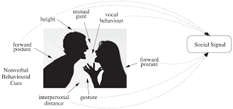 International Workshop on Social Signal Processing and Beyond
**********************************************************************
http://www.ub.edu/cvub/SSPandBE/index.html
September 11, 2017, Catania, Italy
in association with ICIAP 2017 (http://www.iciap2017.com/)
International Workshop on Social Signal Processing and Beyond
**********************************************************************
http://www.ub.edu/cvub/SSPandBE/index.html
September 11, 2017, Catania, Italy
in association with ICIAP 2017 (http://www.iciap2017.com/) Given the increasing quantities of personal data being gathered by individuals, the concept of a digital library of rich multimedia and sensory content for every individual is becoming a reality and fast becoming a mainstream topic for multimedia research. This is often referred to as lifelogging and there are significant challenges to be addressed in the area, concerning the gathering, enriching, searching and accessing of lifelog data.
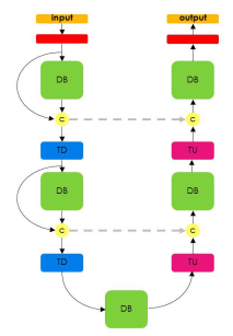 In recent years, deep learning has achieved remarkable results in fields such as: computer vision, speech recognition and natural language processing. This DL revolution is slowly reaching the challenging problems of the medical domain, opening the doors for personalized medicine. Medical domain is characterized by high variability of data including text, imaging, and genomic data. In this talk, we will present recent advances in two domains of medical data: imaging and genomics.
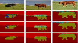
First, we will introduce a simple, yet powerful pipeline for medical image segmentation that combines Fully Convolutional Networks (FCNs) with Fully Convolutional Residual Networks (FC-ResNets). We propose and examine a design that takes particular advantage of recent advances in the understanding of both Convolutional Neural Networks as well as ResNets. Our approach focuses upon the importance of a trainable pre-processing when using FC-ResNets and we show that a low-capacity FCN model can serve as a pre-processor to normalize medical input data. We show that using this pipeline, we exhibit state-of-the-art performance on the challenging Electron Microscopy benchmark, when compared to other 2D methods. We improve segmentation results on CT images of liver lesions, when contrasting with standard FCN methods. Moreover, when applying our 2D pipeline on a challenging 3D MRI prostate segmentation challenge we reach results that are competitive even when compared to 3D methods. The obtained results illustrate the strong potential and versatility of the pipeline by achieving highly accurate results on multi-modality images from different anatomical regions and organs.
Second, we will introduce a novel deep learning architecture to address the challenges posed by genomic data, where the number of input features can be orders of magnitude larger than the number of training examples, making it difficult to avoid overfitting, even when using the known regularization techniques. Improving the ability of deep learning to handle such datasets could have an important impact in medical research, more specifically in precision medicine, where high-dimensional data regarding a particular patient is used to make predictions of interest. We propose a novel neural network parameterization, that we call Diet Networks, which considerably reduces the number of free parameters in the model. The Diet Networks parametrization is based on the idea that we can first learn or provide an embedding for each input feature and then learn how to map a feature's representation to the parameters linking the value of the feature to each of the hidden units of the classifier network. We experiment on a population stratification task of interest to medical studies and show that the proposed approach can significantly reduce both the number of parameters and the error rate of the classifier. This work was accepted at ICLR 2017.
In recent years, deep learning has achieved remarkable results in fields such as: computer vision, speech recognition and natural language processing. This DL revolution is slowly reaching the challenging problems of the medical domain, opening the doors for personalized medicine. Medical domain is characterized by high variability of data including text, imaging, and genomic data. In this talk, we will present recent advances in two domains of medical data: imaging and genomics.
First, we will introduce a simple, yet powerful pipeline for medical image segmentation that combines Fully Convolutional Networks (FCNs) with Fully Convolutional Residual Networks (FC-ResNets). We propose and examine a design that takes particular advantage of recent advances in the understanding of both Convolutional Neural Networks as well as ResNets. Our approach focuses upon the importance of a trainable pre-processing when using FC-ResNets and we show that a low-capacity FCN model can serve as a pre-processor to normalize medical input data. We show that using this pipeline, we exhibit state-of-the-art performance on the challenging Electron Microscopy benchmark, when compared to other 2D methods. We improve segmentation results on CT images of liver lesions, when contrasting with standard FCN methods. Moreover, when applying our 2D pipeline on a challenging 3D MRI prostate segmentation challenge we reach results that are competitive even when compared to 3D methods. The obtained results illustrate the strong potential and versatility of the pipeline by achieving highly accurate results on multi-modality images from different anatomical regions and organs.
Second, we will introduce a novel deep learning architecture to address the challenges posed by genomic data, where the number of input features can be orders of magnitude larger than the number of training examples, making it difficult to avoid overfitting, even when using the known regularization techniques. Improving the ability of deep learning to handle such datasets could have an important impact in medical research, more specifically in precision medicine, where high-dimensional data regarding a particular patient is used to make predictions of interest. We propose a novel neural network parameterization, that we call Diet Networks, which considerably reduces the number of free parameters in the model. The Diet Networks parametrization is based on the idea that we can first learn or provide an embedding for each input feature and then learn how to map a feature's representation to the parameters linking the value of the feature to each of the hidden units of the classifier network. We experiment on a population stratification task of interest to medical studies and show that the proposed approach can significantly reduce both the number of parameters and the error rate of the classifier. This work was accepted at ICLR 2017. Open Postdoctoral fellowship/s in the Dept. de Matemàtiques i Informàtica at Universitat de Barcelona.
The Dept. de Matemàtiques i Informàtica (Mathematics and Computer Science) is looking for and willing to support excellent postdoctoral researchers in the fields of Machine Learning, Computer Vision and Human-Computer Interaction who are interested in applying for a Beatriu de Pinós (BP) 2016 fellowship so as to conduct a two-year postdoc at Universitat de Barcelona.
The purpose of the Beatriu de Pinós programme is to award 60 individuals grants for the hiring and incorporation of postdoctoral research staff into the Catalan science and technology system. These grants are designed for the incorporation of young researchers (who obtained their PhD between 2007 and 2014 and have not resided or worked in Spain for more than 12 months in the three years prior to date of submission of the application), so that they can improve their professional prospects and obtain an independent research position. Candidates must carry out a research and training project for the entire period of the grant, one that will allow them to progress in the development of their professional careers. Please check the website of the BP programme* for further information about this fellowship.
Some of the specific projects, we are working, include:
+ Machine Learning: Deep Learning for time series analysis, Supervised Online Learning Algorithms, Bayesian statistics and deep learning.
+ Computer Vision: Visual Lifelogging and Egocentric Vision, Neuroimage processing, Computer Vision for Food Analysis, Deep learning and Image Aesthetics, Ultrasound image analysis.
+ Human-Computer Interaction: ageing / older people, interfaces for people with mild dementia or with aphasia, universal design of STEM documents
Deadline: 0112/2016.
For further information about this postdoctoral opportunity please feel free to contact us:
Petia Radeva (petia.ivanova@ub.edu or radevap@gmail.com),
www.ub.edu/cvub,
www.cvc.uab.es/people/petia
Open Postdoctoral fellowship/s in the Dept. de Matemàtiques i Informàtica at Universitat de Barcelona.
The Dept. de Matemàtiques i Informàtica (Mathematics and Computer Science) is looking for and willing to support excellent postdoctoral researchers in the fields of Machine Learning, Computer Vision and Human-Computer Interaction who are interested in applying for a Beatriu de Pinós (BP) 2016 fellowship so as to conduct a two-year postdoc at Universitat de Barcelona.
The purpose of the Beatriu de Pinós programme is to award 60 individuals grants for the hiring and incorporation of postdoctoral research staff into the Catalan science and technology system. These grants are designed for the incorporation of young researchers (who obtained their PhD between 2007 and 2014 and have not resided or worked in Spain for more than 12 months in the three years prior to date of submission of the application), so that they can improve their professional prospects and obtain an independent research position. Candidates must carry out a research and training project for the entire period of the grant, one that will allow them to progress in the development of their professional careers. Please check the website of the BP programme* for further information about this fellowship.
Some of the specific projects, we are working, include:
+ Machine Learning: Deep Learning for time series analysis, Supervised Online Learning Algorithms, Bayesian statistics and deep learning.
+ Computer Vision: Visual Lifelogging and Egocentric Vision, Neuroimage processing, Computer Vision for Food Analysis, Deep learning and Image Aesthetics, Ultrasound image analysis.
+ Human-Computer Interaction: ageing / older people, interfaces for people with mild dementia or with aphasia, universal design of STEM documents
Deadline: 0112/2016.
For further information about this postdoctoral opportunity please feel free to contact us:
Petia Radeva (petia.ivanova@ub.edu or radevap@gmail.com),
www.ub.edu/cvub,
www.cvc.uab.es/people/petia
 Petia Radeva has been invited speaker to the workshop “Humanitarian and social science: from the university to the enterprise” 17 of November, 2016, Faculty of Filology, University of Barcelona.
 Best paper award at CIAPR'2016 to our paper: "Deep Learning Features for Wireless Capsule Endoscopy Analysis",
by Santi Segui, Michal Drozdzal, Guillem Pascual, Carolina Malagelada, Fernando Azpiroz, Petia
Radeva and Jordi Vitrià, Lima Perú, 2016.
Best paper award at CIAPR'2016 to our paper: "Deep Learning Features for Wireless Capsule Endoscopy Analysis",
by Santi Segui, Michal Drozdzal, Guillem Pascual, Carolina Malagelada, Fernando Azpiroz, Petia
Radeva and Jordi Vitrià, Lima Perú, 2016. Petia Radeva received the International CIARP Award "Aurora Pons Porrata" in recognition of an outstanding technical contribution to the field of pattern recognition, data mining and related areas, 2016.
 We got the Mention prize (II place) in the DKV competence with our App for Automatic Food Recognition for Healthy Habits Promotion! Congratulations, team!!!
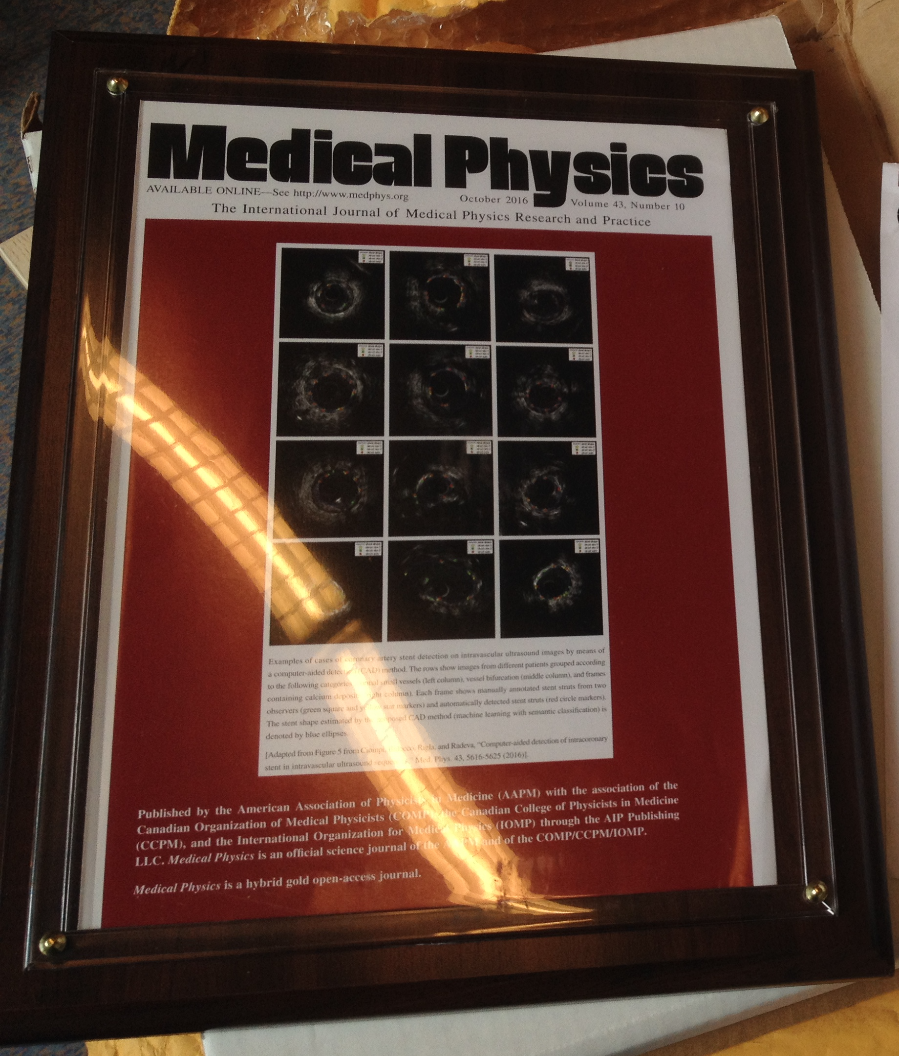 The journal Medical Physics chose figures of our work on Stent analysis to use as a cover on their journal. http://www.medphys.org
 Petia Radeva gave a plenary talk "Can Deep Learning and Egocentric Vision for Visual Lifelogging help us eat better?" at the CCIA'2016, organized by Xavier Binefa, UPF, Barcelona, Spain.
 The prestigous GRADIANT award was assifned to Beatriz Remeseiro for the best PhD thesis applied to the ICT sector 2016.
 3 abstracts accepted at the NIPS Workshop WiML'16. Congratulations, Mariella, Maya and Beatriz!
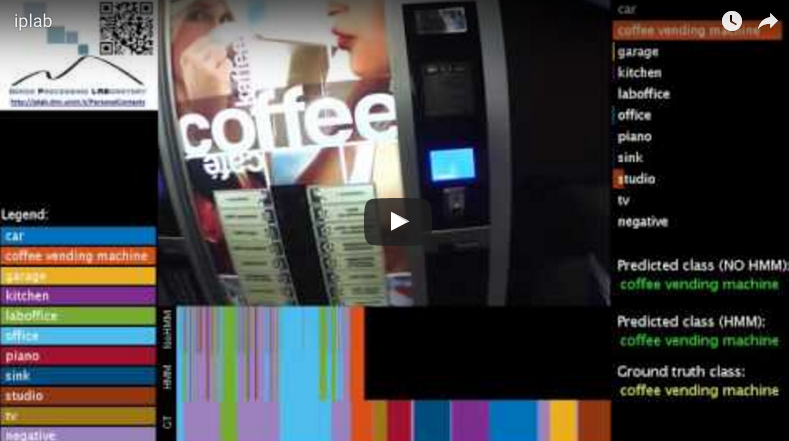
 On 8 of February, 2016
  Michal Drozdzal is one of the 5 researchers who received the Pioneer Award for her doctoral thesis "Sequential image analysis for computer-aided endoscopi wireless". This is the second edition of the competition promoted by CERCA. This year a total of nineteen researchers (10 males and 9 females) participated from thirteen centres nearby. By areas, there have been 3 science projects; 7 of medical sciences and health; 3 of 6 engineering and life sciences. The jury appreciated the advanced technology that solves problems linked to the endoscopy with a significant population component and a business link which enables a good transfer of knowledge generated.
 Re-memory: Cognitive Training based on autobiograohic records to exercise memory
Exposition “+HUMANS”, 17 December 2015, CCB, Barcelona Spain
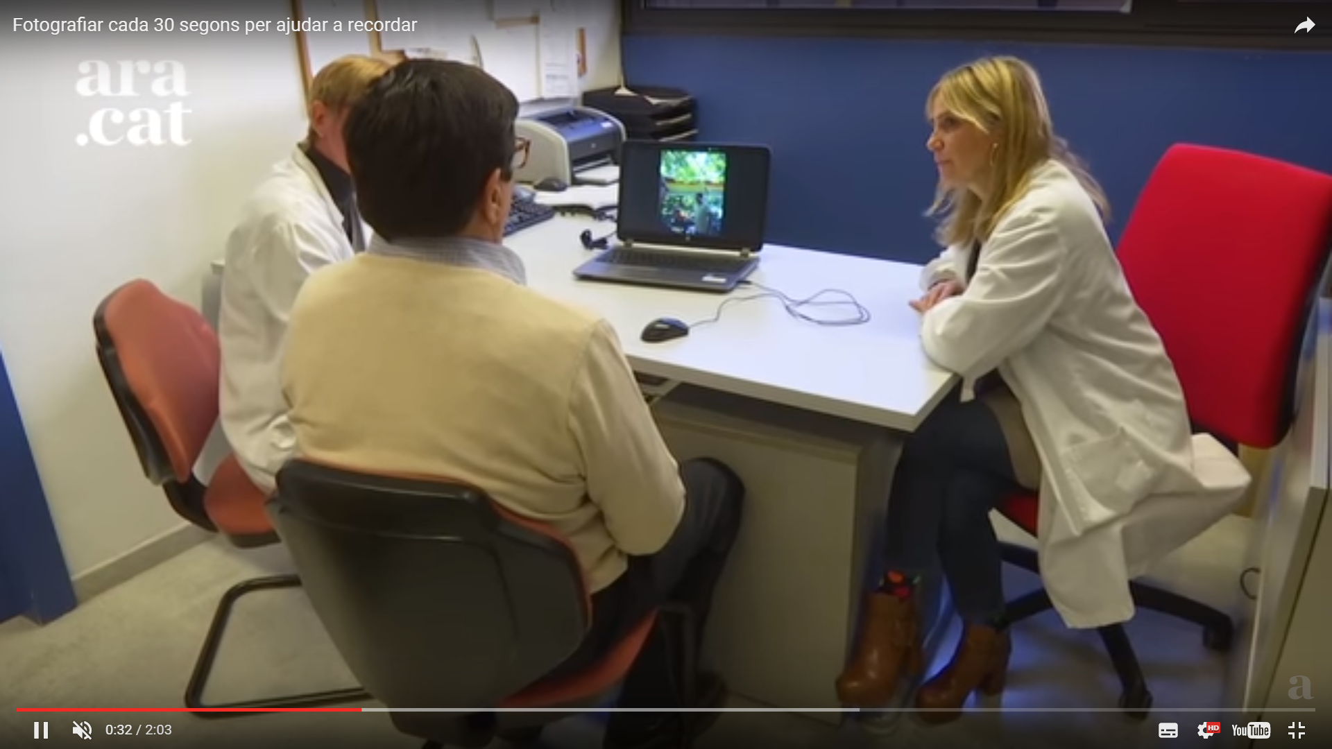
The majority of patients with mild cognitive impairment develop dementia and Alzheimer's disease. Re-memory, a project financed by the Foudation “La Marató de TV3”, study a new entrenament cognitiu based on the concept of lifelogging to exrcise memory. Re-memory works in the following way: the patient carries a camera that capture automatically images from all locations visited, events in which the wearer participates, the activities that he/she participated and persons he/she interacted with. Inspired in the use of wearable cameras for first cases of amnesia, els membres of the Re-memory project create a cogntive program for training based on the autobiographic reexperimenting to positively impact the cognition and improve the memory and function of peoplewith mild cognitive impairment.
Re-memory: Cognitive Training based on autobiograohic records to exercise memory
Exposition “+HUMANS”, 17 December 2015, CCB, Barcelona Spain
The majority of patients with mild cognitive impairment develop dementia and Alzheimer's disease. Re-memory, a project financed by the Foudation “La Marató de TV3”, study a new entrenament cognitiu based on the concept of lifelogging to exrcise memory. Re-memory works in the following way: the patient carries a camera that capture automatically images from all locations visited, events in which the wearer participates, the activities that he/she participated and persons he/she interacted with. Inspired in the use of wearable cameras for first cases of amnesia, els membres of the Re-memory project create a cogntive program for training based on the autobiographic reexperimenting to positively impact the cognition and improve the memory and function of peoplewith mild cognitive impairment.
 Our work on using Deep Learning techniques for endoluminal image analysis was presented as example at NVIDIA’s GPU Technical Conference (http://www.gputechconf.com/).