Association of TGF-beta1 polymorphisms with
chronic renal disease
E. Coll1, B. Cormand2, B.
Campos3, D. González-Núñez4, P. Iñigo5, A.
Botey1, E. Poch1
1Nephrology Service, Hospital Clinic, Faculty of Biology,
Faculty of Medicine, University of Barcelona - Spain
2Department of Genetics, Faculty
of Biology, University of Barcelona - Spain 3Unit of Biostatistics, Faculty of Medicine, University of
Barcelona, Barcelona - Spain 4Nephrology Service, Hospital Clinic, Faculty of Biology,
Faculty of Medicine, University of Barcelona - Spain and Department of Genetics,
Faculty of Biology, University of Barcelona - Spain 5Laboratorio de Hormonal IDIBAPS, Hospital Clinic, Barcelona -
Spain
ABSTRACT:Background: Transforming
growth factor beta1 (TGF-ß1) plays an important role in tissue fibrosis and has
been found to participate in cardiovascular disease (CVD). This study aimed to
evaluate the association of TGF-ß1 polymorphisms with chronic renal disease
(CRD), and its progression to dialysis in a retrospective longitudinal study of
an end-stage renal disease (ESRD) cohort. Methods: The Arg/Pro (codon
25) and Leu/Pro (codon 10) polymorphisms were genotyped in 104 ESRD patients
aged 64 ± 14 yrs (mean ± SD), 62 males, and in 104 matched controls.
Results: The genotype distribution of Leu10Pro and Arg25Pro
polymorphisms was different between patients and controls: Leu/Leu, Leu/Pro,
Pro/Pro: 0.35, 0.50, 0.15 vs. 0.30, 0.24, 0.46 (p=0.001) and Arg/Arg, Arg/Pro,
Pro/Pro: 0.79, 0.21, 0 vs. 0.87, 0.10, 0.03 (p=0.019). Similarly, haplotypes
constructed with the combination of both polymorphisms were different among
groups. There were no differences in CRD progression rate among genotypes. Codon
10 Leu allele was associated with the presence of clinical CVD in the ESRD
patients (Leu/Leu, Leu/Pro, Pro/Pro: with CVD 0.49, 0.49, 0.02 vs. without CVD
0.27, 0.51, 0.22 (p=0.01). Combined polymorphism haplotypes were also
significantly different between ESRD patients with and without CVD. This
association was independent from other risk factors. Conclusions:
TGF-ß1 polymorphisms are associated with ESRD, particularly in patients with
associated clinical CVD, and could be useful as genetic markers of CRD and
higher cardiovascular risk.
Chronic renal
disease (CRD), once established, tends to progress to end-stage renal disease
(ESRD). Tissue fibrosis is the histological hallmark of progressive renal
disease, and is relatively independent from the primary etiology (1). On the
other hand, patients with progressive CRD or ESRD have an exceedingly high risk
of cardiovascular morbidity and mortality, which is not fully explained by
traditional risk factors (2). The renin-angiotensin-aldosterone system has a
fund-amental role in progressive CRD development (3, 4) and possibly its
complications, but recent attention has centered on downstream cytokines as
potential participants and future therapeutic targets. It is known that the
pro-hypertrophic actions of angiotensin II are mediated by growth factors (5).
Transforming growth factor beta1 (TGF-ß1) is a multifunctional cytokine that
regulates cell growth, differentiation and matrix production, inducing fibrosis
in a variety of tissues such as kidney, heart and blood vessels (6).
Specifically, TGF-ß1 overproduction has been linked to target organ damage in
hypertension (7), and TGF-b1 gene transfer in the mesangium of normal rats leads
to glomerulosclerosis (8). In human disease, increased TGF-ß production has been
described in nephropathies such as glomerulonephritis, diabetic nephropathy,
nephroangiosclerosis and allograft nephropathy (9). The production and secretion
of TGF-ß1 in humans seem to be, in part, genetically regulated (10). Eight
TGF-ß1 polymorphisms have recently been reported, the (Leu 10→Pro and Arg
25→Pro) being the most frequently studied for their association to disease and
to the rate of cytokine production (11-14). Associations between TGF-ß1 gene
polymorphisms and cardiovascular disease (CVD) such as myocardial infarction,
diabetic nephropathy, hypertension and serum TGF-ß1 levels (11, 13-15) have been
described. This study aimed to analyze the association of the Leu 10→Pro and
Arg 25→Pro TGF-ß1 polymorphisms with advanced CRD, CRD progression rate, and
with the clinical characteristics and cardiovascular risk factors of this
high-risk population.
Subjects and methods
Patients and clinical data
We studied 104
Caucasian ESRD patients on hemodialysis (HD) from the Division of Nephrology
(Hospital Clinic, Nephrology Service, University of Barcelona), and 104
Caucasian controls. Clinical information and biochemical parameters were
retrieved retrospectively from hospital records. Patients were selected with at
least four serum creatinine (Cr) measurements spanning a minimum 1 yr of
follow-up before HD start to calculate the slope of reciprocal Cr vs. time (1).
Risk factors for CVD and for CRD progression at the time of renal insufficiency
diagnosis were recorded: age, sex, present or past cigarette smoking,
hypertension (blood pressure (BP) ≥140 or 90 mmHg), diabetes mellitus (fasting
blood glucose ≥126 mg/dL), and dyslipidemia (serum total cholesterol >240
mg/dL and/or serum triglycerides >200 mg/dL). Clinical CVD was defined as the
presence of at least one episode of the following (1) cerebrovascular accident
documented by CT scan, (2) coronary artery disease defined as anginal episodes
or myocardial infarction based on ECG changes, serum enzymes or angiography and;
(3) peripheric vasculopathy defined by intermittent claudication or occlusive
disease documented by angiography. The ethical committee of the Hospital Clinic
approved the study. Consent was obtained from all patients for inclusion in the
study. As a control group, 104 subjects from the same hospital admitted for
elective surgery were selected with the following inclusion criteria: (1) age
between 25 and 85 yrs, (2) absence of nephropathy or renal failure, diabetes
mellitus or CVD.
Molecular studies
Genomic DNA was
isolated from peripheral-blood lymphocytes by the standard salting-out procedure
(16). The Leu 10→Pro polymorphism (a T→C transition at codon 10) and the Arg
25→Pro polymorphism (a G→C transversion at codon 25) of the TGF-ß1 gene were
genotyped with flanking primers and restriction digestion (MspA1 and FseI,
respectively), as described previously (16).
Statistical
analysis
Results are reported as mean ± SD for normally
distributed continuous variables, median (range) for non normal variables or as
frequencies for categorical variables. Differences between genotype groups were
analyzed using the analysis of variance (ANOVA) test followed by the Bonferroni
multiple comparison or by the χ² test when appropriate. Hardy-Weinberg
equilibrium was tested for each polymorphic site by the χ² test. Linkage
disequilibrium between the two TGF-ß1 polymorphisms analyzed was evaluated using
EH software (17). Haplotype estimations from the population genotype data were
performed using PHASE version 2.0 software (18). This program was also used to
perform a case-control permutation test for significant differences in haplotype
frequencies in patient and control groups: ESRD vs. controls, ESRD with CVD vs.
ESRD without CVD. Logistic regression analysis was performed to identify the
factors independently associated with cardiovascular complications in CRD
patients. Variables were included when a value p<0.3 was obtained in
univariate analysis with CVD as a dependent variable and clinical and
biochemical variables as independent variables. CRD progression to dialysis was
evaluated in each patient as renal function loss by the slope of 1/Cr vs. time.
As the distribution of the slopes was skewed, log transformation, ln (-1x1/Cr),
were applied to yield more normally distributed data. Statistical tests were
two-tailed, and p<0.05 was used to identify statistically significant
results. Computer analyzes were done using SSPS-PC software version 11 (SPSS
Inc).
Results
The ESRD patients included 104 Caucasians,
62 males and 42 females aged 64 ± 14 yrs (range 25-89 yrs) with systolic blood
pressure (SBP) of 157 ± 20 mmHg and diastolic blood pressure (DBP) of 87 ± 9
mmHg and a plasma Cr at presentation of 2.6 ± 1.1 mg/dL. The median
follow-up period was 51.6 months (range 12-244). The etiology categories for CRD
were nephro-angiosclerosis (n=35), diabetes mellitus (n=21), specified and
unspecified glomerulonephritis (n=16), autosomal-dominant polycystic kidney
disease (n=10), interstitial nephritis (n=5) and undetermined cause (n=19).
Clinical CVD was present in 37 patients; cerebrovascular disease (n=10),
ischemic heart disease (n=24) and peripheric vascular disease (n=14). A control
group consisted of 104 Caucasians; 58 males and 46 females aged 60 ± 13 yrs with
a mean SPB and mean DBP of 117 ± 11 and 69 ± 7 mmHg, respectively. There were no
significant differences in age or gender distribution between patients and
controls.
DISTRIBUTION OF GENOTYPES AMONG PATIENTS AND
CONTROLS
TABLE
I
The frequencies of the different genotypes studied did not
deviate from the Hardy-Weinberg equilibrium in patients and in controls except
for Leu10Pro in controls. As shown in Table I, the genotype distribution between
ESRD patients and controls was different for both the Arg25Pro polymorphism and
the Leu10Pro polymorphism. When haplotypes combining both sites were
constructed, the Leu10+Arg25 haplotype was shown to be overrepresented in the
ESRD group when compared to controls (p=0.001) (Tab. II) by using the
permutation test (PHASE version 2.0). Similarly, there was a significant
difference in haplotype distribution among patients and controls when using the
χ² test (χ²=32.46, 6 d.f., p<0.001). The two polymorphisms were not in
linkage disequilibrium in the ESRD patients, the controls or the complete sample
set, indicating that the variants appeared to be randomly associated.
DISTRIBUTION OF RECONSTRUCTED HAPLOTYPES OF THE
LEU10PRO AND ARG25PRO TGF-b1 POLYMORPHISMS IN ESRD CASES, HEALTHY
CONTROLS, ESRD CASES WITH CVD AND ESRD CASES WITHOUT CVD
TABLE
II
CLINICAL AND BIOCHEMICAL CHARACTERISTICS OF THE
PATIENTS AMONG GENOTYPES OF TGF-b1 CODON 10 AND CODON 25 POLYMORPHISMS
TABLE
III
Table III reports the clinical characteristics of the patients
regarding the TGF-ß1 polymorphism genotypes. There were no differences in age,
gender, BP, smoking status, dyslipidemia or diabetes among genotypes. The
relationship between the TGF-ß1 polymorphisms and CRD progression rate was
analyzed and, as shown in Table III, no differences were found. Regarding the
clinical covariates, the Leu allele of codon 10 polymorphism was significantly
associated with the presence of clinical CVD. Haplotype analysis showed that the
Leu10+Arg25 combination was overrepresented in ESRD patients with CVD when
compared to ESRD patients without CVD (p=0.011) (Tab. II) by using the
permutation test. The χ² test confirmed these results (χ²=11.88, 5 d.f.,
p=0.036).
CLINICAL CHARACTERISTICS OF THE ESRD PATIENTS
ACCORDING TO THE PRESENCE OR ABSENCE OF CLINICAL CVD
TABLE IV
Table IV shows the univariate analysis of CVD risk fact-ors
as independent variables and the presence or absence of CVD as dependent
variables. In addition to codon 10 polymorphism, age, dyslipidemia and diabetes
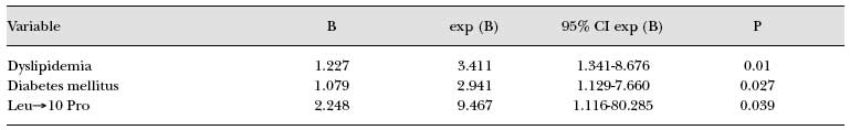
were associated with clinical CVD in the ESRD patients. A logistic regression
analysis indicated that the association between the codon 10 polymorphism and
clinical CVD was independent. Indeed, only dyslipidemia (p=0.010), diabetes
(p=0.027) and codon 10 polymorphism (p=0.039) entered the logistic regression
model as independent predictors of CVD in ESRD patients (Tab.
V).
POINTWISE AND 95% CONFIDENCE INTERVAL ESTIMATES
OF THE REGRESSION COEFFICIENTS FOR THE LOGISTIC REGRESSION MODEL
TABLE V
Discussion
We analyzed the association of two
TGF-ß1 polymorphisms with ESRD and CRD progression. We observed an association
between the Arg25Pro and Leu10Pro polymorphisms with CRD. This association was
also significant when haplotypes combining both sites were constructed
(Leu10+Arg25 haplotype). It is important to note that the two polymorphisms were
not in linkage disequilibrium in the ESRD group, the controls or the complete
sample set; therefore, indicating that the variants appeared to be randomly
associated and supplied independent information. In addition, the Leu10Pro
polymorphism was significantly and independently associated with the presence of
CVD in the ESRD patients studied. Similarly, haplotype analysis showed that the
Leu10+Arg25 combination was overrepresented in ESRD patients with CVD when
compared to ESRD patients without CVD. Variants of the TGF-ß1 gene have
previously been associated with differences in the production, secretion or
activity of this cytokine (13). Through several mechanisms, the variable
availability of this growth factor in different tissues could affect endothelial
function (19, 20) and influence BP, interfere with the development of
atherosclerosis (21), and influence vascular (22-24) and cardiac (25)
remodeling. The independent association between the Leu10Pro polymorphism
and the presence of clinical CVD in ESRD patients could be clinically important
indicating its potential use as a genetic marker for higher cardiovascular risk
in ESRD patients. It is known that ESRD patients have a very high risk of CVD,
which is not fully explained by the presence or accumulation of traditional risk
factors (2). TGF-ß1 polymorphisms have been associated with myocardial
infarction, hypertension and serum TGF-ß1 levels (11, 14, 26) in non-renal
patients. In the ECTIM STUDY (11), the Pro25 allele was associated with an
increased risk of myocardial infarction and a reduced risk of hypertension.
Similarly, Li et al (14) found that the Arg25 allele was more frequent in
hypertensive subjects. Recently, Yokota et al (26) described an association
between the Leu10 allele and susceptibility to myocardial infarction in males
with conventional cardiovascular risk factors. In agreement with the latter
study, we observed an association between clinical CVD and the Leu10 allele. The
important difference is that this association was also present in CRD patients,
this study being the first to explore this issue in this type of high-risk CVD
patients. Serum TGF-ß levels were not measured in this retrospective study. It
is known that present serum TGF-ß levels would not be useful as predictor
variables or for pathogenic associations, since these levels are influenced by
disease status (26). Finally, concerning CRD and progression, we found that
TGF-ß1 gene variants did not influence the progression rate. This is
controversial since Pociot et al (15) found a weak but significant association
of the Thr263Ile variant with diabetic nephropathy, whereas Akai et al (27) did
not find an association between the codon 10 Leu/Leu genotype and faster
progression in this disease. Recent work has shown a correlation between the
TGF-ß1 Leu10 allele and more severe histological renal damage and with
progressive renal function deterioration in IgA nephropathy (28, 29). The
finding that allele Leu10 was more frequent in ESRD patients than in controls
correlates with the results observed by other studies (29) and is consistent
with the idea that TGF-ß could participate as a key factor in the common
mechanisms leading to tissue fibrosis and the development of advanced CRD of
various etiologies. In conclusion, TGF-ß1 polymorphisms are associated with
CRD and could be markers for higher cardiovascular risk in this population.
Tissue and vascular growth and fibrosis are common mechanisms for the
development of renal and cardiovascular disease and these results suggest that
TGF-ß1 gene variants could play a role in both processes. However, due to the
common problems concerning the lack of reproducibility of genetic association
studies, probably resulting from a mix of type 2 errors, sample stratifications
and inadequate sample size (30), larger and prospective studies are necessary to
confirm these results.
Acknowledgements
This work was
supported in part by a grant from the Fondo de Investigaciones Sanitarias, FIS
01/1151, and a grant by the Hospital Clinic (to E.C.).
Address
for correspondence: Esteban Poch, M.D. Servicio de Nefrología
Hospital Clínic Universidad de Barcelona Villarroel 170 08036
Barcelona, Spain epoch@medicina.ub.es
REFERENCES(when available, each reference has been linked to
PubMed)
Received: April 15, 2004
Revised: August 06, 2004 Accepted: October 11, 2004